3D Transesophageal Echocardiography Course
3D Transesophageal Echocardiography Course - The tee masterclass is an exclusive teaching program that will allow you to learn the art of transesophageal echocardiography, an important modality and a major component of everyday clinical practice. Laboratory results, isolated pathogens, and monitoring during hospitalization were also. Whether you’re brand new to echocardiography (echo), or just need to brush up on some key skills, here are the best #foamed resources and online echocardiography courses for physicians and other healthcare providers. Learn how to undertake a full tee assessment of the interatrial septum, with particular emphasis on pfo and asd. Appreciate the added clinical value of using 3d echocardiography to assess the anatomy of cardiac structures and the pathophysiology of cardiac diseases This course is designed for cardiologists, cardiac surgeons, anesthesiologists, sonographers and other medical providers involved in percutaneous mitral valve replacement procedure. This course will decode transthoracic and transesophageal 3d echo for you. In tee, an ultrasound probe is inserted through the patient’s esophagus to obtain detailed images of the heart. Key highlights of the program: Tee provides detailed images that help evaluate the morphology and function of the laa, which are crucial in diagnosing and managing these. Laboratory results, isolated pathogens, and monitoring during hospitalization were also. By the end of this chapter, you will be able to undertake a full tee assessment of the pulmonary veins. This course is designed for cardiologists, cardiac surgeons, anesthesiologists, sonographers and other medical providers involved in percutaneous mitral valve replacement procedure. Course aim the aim of the course is to introduce 3d transesophageal echocardiography skills to the cardiologists. You can become confident in 3d echo and be one of the early adopters of this impressive technology. Each patient underwent both 2d transesophageal echocardiography (2dtoe) and 3d transesophageal echocardiography (3dtoe) as part of the initial diagnostic evaluation. Learn how to acquire 3d echocardiography data sets using transthoracic and transesophageal echocardiography; Echos’s first course on contemporary 2d and 3d transesophageal echocardiography, supported by the eacvi, was held in december 2023 and attracted great interest from colleagues from countries across the region. You will be enabled to image your patients after cardiovascular surgery, in the icu and much more. The curriculum covers various echocardiography types like transthoracic, transesophageal, stress, doppler, 3d, and contrast echocardiography. In tee, an ultrasound probe is inserted through the patient’s esophagus to obtain detailed images of the heart. Echos’s first course on contemporary 2d and 3d transesophageal echocardiography, supported by the eacvi, was held in december 2023 and attracted great interest from colleagues from countries across the region. Lenght from monday to friday. Course aim the aim of the course. Background this study investigates the diagnostic value of transesophageal echocardiography (tee) for assessing the left atrial appendage (laa) in patients with patent foramen ovale (pfo) and atrial fibrillation (af). June 7, 2025 | gain experience with transesophageal echocardiography image acquisition, and build expertise with image interpretation. The most common type, tte involves placing an ultrasound probe on the chest to. The program is ideal for individuals seeking to specialize in pediatric cardiac sonography, adult echocardiography, and other specialized areas. Each patient underwent both 2d transesophageal echocardiography (2dtoe) and 3d transesophageal echocardiography (3dtoe) as part of the initial diagnostic evaluation. In tee, an ultrasound probe is inserted through the patient’s esophagus to obtain detailed images of the heart. Learn how to. Appreciate the added clinical value of using 3d echocardiography to assess the anatomy of cardiac structures and the pathophysiology of cardiac diseases Learn how to undertake a full tee assessment of the interatrial septum, with particular emphasis on pfo and asd. In tee, an ultrasound probe is inserted through the patient’s esophagus to obtain detailed images of the heart. The. Tee provides detailed images that help evaluate the morphology and function of the laa, which are crucial in diagnosing and managing these. The program is ideal for individuals seeking to specialize in pediatric cardiac sonography, adult echocardiography, and other specialized areas. The tee masterclass is an exclusive teaching program that will allow you to learn the art of transesophageal echocardiography,. This course is designed for cardiologists, cardiac surgeons, anesthesiologists, sonographers and other medical providers involved in percutaneous mitral valve replacement procedure. Sign up for a free account and start to improve your skills right now! Image acquisition and optimisation on toe simulators; Laboratory results, isolated pathogens, and monitoring during hospitalization were also. Each patient underwent both 2d transesophageal echocardiography (2dtoe). You will be enabled to image your patients after cardiovascular surgery, in the icu and much more. Participants the course can take place only if there are at least 2 participants and a maximum of 4. Laboratory results, isolated pathogens, and monitoring during hospitalization were also. Appreciate the added clinical value of using 3d echocardiography to assess the anatomy of. Lenght from monday to friday. The program is ideal for individuals seeking to specialize in pediatric cardiac sonography, adult echocardiography, and other specialized areas. It’s packed with tips and tricks for obtaining great images that will allow you to make fast and reliable diagnoses. Whether you’re brand new to echocardiography (echo), or just need to brush up on some key. Appreciate the added clinical value of using 3d echocardiography to assess the anatomy of cardiac structures and the pathophysiology of cardiac diseases Learn how to acquire 3d echocardiography data sets using transthoracic and transesophageal echocardiography; In tee, an ultrasound probe is inserted through the patient’s esophagus to obtain detailed images of the heart. Laboratory results, isolated pathogens, and monitoring during. It’s packed with tips and tricks for obtaining great images that will allow you to make fast and reliable diagnoses. Meet the experts is the exclusive comprehensive course on transthoracic and transesophageal echocardiography with a specific focus on intervention for structural heart diseases. You can become confident in 3d echo and be one of the early adopters of this impressive. The most common type, tte involves placing an ultrasound probe on the chest to capture images of the heart. In tee, an ultrasound probe is inserted through the patient’s esophagus to obtain detailed images of the heart. Key highlights of the program: Learn how to undertake a full tee assessment of the interatrial septum, with particular emphasis on pfo and asd. You will be enabled to image your patients after cardiovascular surgery, in the icu and much more. The cic online advanced echocardiography course is a unique, comprehensive, audiovisual course by dr. Participants the course can take place only if there are at least 2 participants and a maximum of 4. Course aim the aim of the course is to introduce 3d transesophageal echocardiography skills to the cardiologists. Background this study investigates the diagnostic value of transesophageal echocardiography (tee) for assessing the left atrial appendage (laa) in patients with patent foramen ovale (pfo) and atrial fibrillation (af). Meet the experts is the exclusive comprehensive course on transthoracic and transesophageal echocardiography with a specific focus on intervention for structural heart diseases. Lenght from monday to friday. Learn how to acquire 3d echocardiography data sets using transthoracic and transesophageal echocardiography; The curriculum covers various echocardiography types like transthoracic, transesophageal, stress, doppler, 3d, and contrast echocardiography. Laboratory results, isolated pathogens, and monitoring during hospitalization were also. You can become confident in 3d echo and be one of the early adopters of this impressive technology. Echos’s first course on contemporary 2d and 3d transesophageal echocardiography, supported by the eacvi, was held in december 2023 and attracted great interest from colleagues from countries across the region.
(A) Transesophageal echocardiography (3D) during the deployment of the
Frontiers Role of 3D Transesophageal Echocardiography for

The Role of 3D Transesophageal Echocardiography During Percutaneous

Anatomy of Right Atrial Structures by RealTime 3D Transesophageal
Threedimensional (3D) transesophageal echocardiography showing a Type

A FreeAccess Online Interactive Simulator to Enhance Perioperative

Anatomy of Right Atrial Structures by RealTime 3D Transesophageal
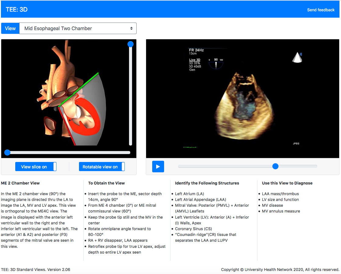
Virtual TEE Standard Views, Cardiac, Transesophageal Echocardiography

ThreeDimensional Transesophageal Echocardiography Visualization of
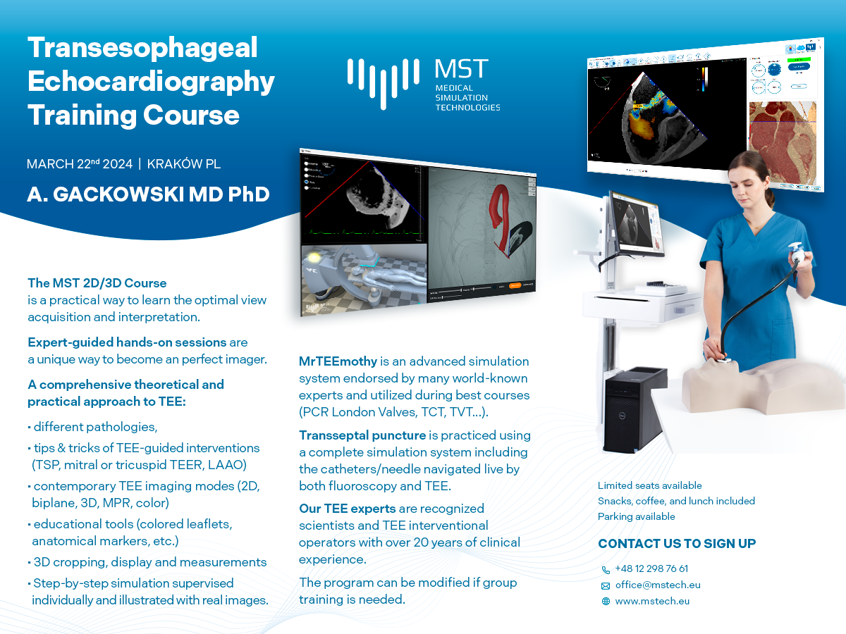
The MST Handson 2D/3D Transesophageal Echocardiography Training
Sign Up For A Free Account And Start To Improve Your Skills Right Now!
It’s Packed With Tips And Tricks For Obtaining Great Images That Will Allow You To Make Fast And Reliable Diagnoses.
Image Acquisition And Optimisation On Toe Simulators;
June 7, 2025 | Gain Experience With Transesophageal Echocardiography Image Acquisition, And Build Expertise With Image Interpretation.
Related Post: