Course Of Ureter In Female Pelvis
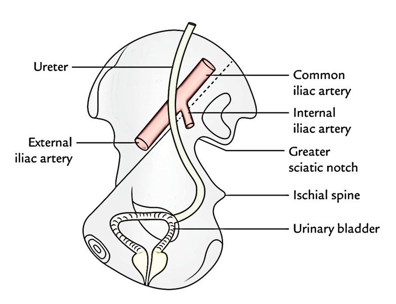
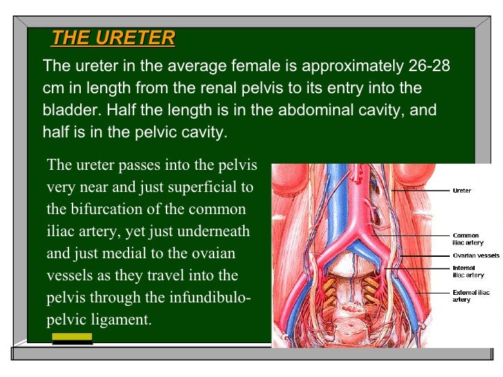
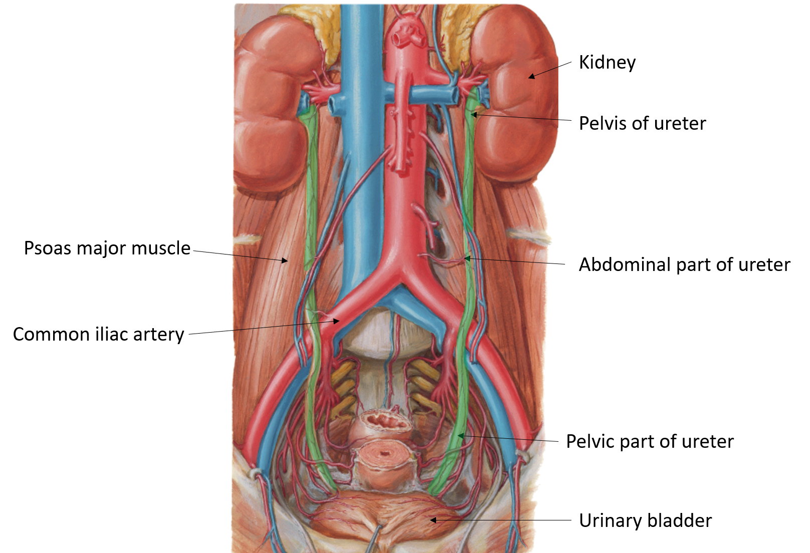
Course Of Ureter In Female Pelvis - During their course in the abdomen, the ureters receive blood from the gonadal vessels, aorta, and retroperitoneal vessels. Radiographic anatomy of the ureter]:. In the majority of the patients, the course of the ureter is easily demarcated from the level of the pelvic brim. From the renal pelvis to the pelvic brim. Within the abdomen, the ureters descend retroperitoneally and anterior to the muscles of the posterior abdominal wall (psoas major), eventually, they reach the pelvic brim where they. In the female, the ureter forms, as it lies in relation to the wall of the pelvis, the posterior boundary of a shallow depression named the ovarian fossa, in which the ovary is situated. From the pelvic brim to the bladder. In females, they sit posterior to the ovary, and then run adjacent to the cervix and lateral fornices of the vagina before entering the bladder. Then, at the level of the ischial spine, the uterine artery crosses the ureter. In the pelvis, they receive additional branches from the internal. Within the abdomen, the ureters descend retroperitoneally and anterior to the muscles of the posterior abdominal wall (psoas major), eventually, they reach the pelvic brim where they. From the renal pelvis to the pelvic brim. In the pelvis, they receive additional branches from the internal. Radiographic anatomy of the ureter]:. Turn downward through wall of pelvis3.cross ureteric canal4. Fast shippingshop best sellersshop our huge selectiondeals of the day So, the ureter has two divisions: In females, they sit posterior to the ovary, and then run adjacent to the cervix and lateral fornices of the vagina before entering the bladder. In the female, the ureter forms, as it lies in relation to the wall of the pelvis, the posterior boundary of a shallow depression named the ovarian fossa, in which the ovary is situated. Thus, if there is any abnormality in. From the pelvic brim to the bladder. In females, the ureters pass medial to the origin of the uterine artery. In the pelvis, they receive additional branches from the internal. On radiographs, the ureter is divided into three sections [fig. Within the abdomen, the ureters descend retroperitoneally and anterior to the muscles of the posterior abdominal wall (psoas major), eventually,. From the renal pelvis to the pelvic brim. Radiographic anatomy of the ureter]:. The urethra is a fibromuscular tube that conducts urine from the bladder (and semen from the ductus deferens) to the exterior. Then, at the level of the ischial spine, the uterine artery crosses the ureter. The ureters regulate the course of the urine, in a single direction. Fast shippingshop best sellersshop our huge selectiondeals of the day Within the abdomen, the ureters descend retroperitoneally and anterior to the muscles of the posterior abdominal wall (psoas major), eventually, they reach the pelvic brim where they. However, they do not work like other body sphincters, preventing reflux. The ureters regulate the course of the urine, in a single direction.. From the renal pelvis to the pelvic brim. During their course in the abdomen, the ureters receive blood from the gonadal vessels, aorta, and retroperitoneal vessels. Thus, if there is any abnormality in. Cross pelvic brim infront of internal iliac artery bifurcation2. In females, they sit posterior to the ovary, and then run adjacent to the cervix and lateral fornices. So, the ureter has two divisions: Finally, the ureters run near the lateral part. Its upper half courses in the abdomen (abdominal part) while its lower half courses in the pelvis (pelvic part). Cross pelvic brim infront of internal iliac artery bifurcation2. In the pelvis, they receive additional branches from the internal. Cross pelvic brim infront of internal iliac artery bifurcation2. Its upper half courses in the abdomen (abdominal part) while its lower half courses in the pelvis (pelvic part). From the renal pelvis to the pelvic brim. In the majority of the patients, the course of the ureter is easily demarcated from the level of the pelvic brim. In general the. The ureters regulate the course of the urine, in a single direction. In general the ureter is seen crossing the external iliac vessels from lateral to. In the female, the ureter forms, as it lies in relation to the wall of the pelvis, the posterior boundary of a shallow depression named the ovarian fossa, in which the ovary is situated.. So, the ureter has two divisions: It begins at the neck of the bladder, traverses the pelvic and. However, they do not work like other body sphincters, preventing reflux. Thus, if there is any abnormality in. On radiographs, the ureter is divided into three sections [fig. In general the ureter is seen crossing the external iliac vessels from lateral to. In females, the ureters pass medial to the origin of the uterine artery. Then, at the level of the ischial spine, the uterine artery crosses the ureter. However, they do not work like other body sphincters, preventing reflux. Its upper half courses in the abdomen (abdominal. The urethra is a fibromuscular tube that conducts urine from the bladder (and semen from the ductus deferens) to the exterior. In the female, the ureter forms, as it lies in relation to the wall of the pelvis, the posterior boundary of a shallow depression named the ovarian fossa, in which the ovary is situated. Within the abdomen, the ureters. From the pelvic brim to the bladder. Thus, if there is any abnormality in. However, they do not work like other body sphincters, preventing reflux. From the renal pelvis to the pelvic brim. In females, they sit posterior to the ovary, and then run adjacent to the cervix and lateral fornices of the vagina before entering the bladder. In the female, the ureter forms, as it lies in relation to the wall of the pelvis, the posterior boundary of a shallow depression named the ovarian fossa, in which the ovary is situated. Cross pelvic brim infront of internal iliac artery bifurcation2. In females, the ureters pass medial to the origin of the uterine artery. Fast shippingshop best sellersshop our huge selectiondeals of the day During their course in the abdomen, the ureters receive blood from the gonadal vessels, aorta, and retroperitoneal vessels. On radiographs, the ureter is divided into three sections [fig. The urethra is a fibromuscular tube that conducts urine from the bladder (and semen from the ductus deferens) to the exterior. Finally, the ureters pass through the bladder wall in. Radiographic anatomy of the ureter]:. Within the abdomen, the ureters descend retroperitoneally and anterior to the muscles of the posterior abdominal wall (psoas major), eventually, they reach the pelvic brim where they. Finally, the ureters run near the lateral part.
Anatomy of the Female Urinary Tract Obgyn Key
Ureter Earth's Lab
Pelvic ureter
Anatomy2009
Course of pelvic ureters. Taken from [1]. Download Scientific Diagram

Cardinal Ligament Ureter

Course of ureter female Diagram Quizlet
Ureter Anatomy QA
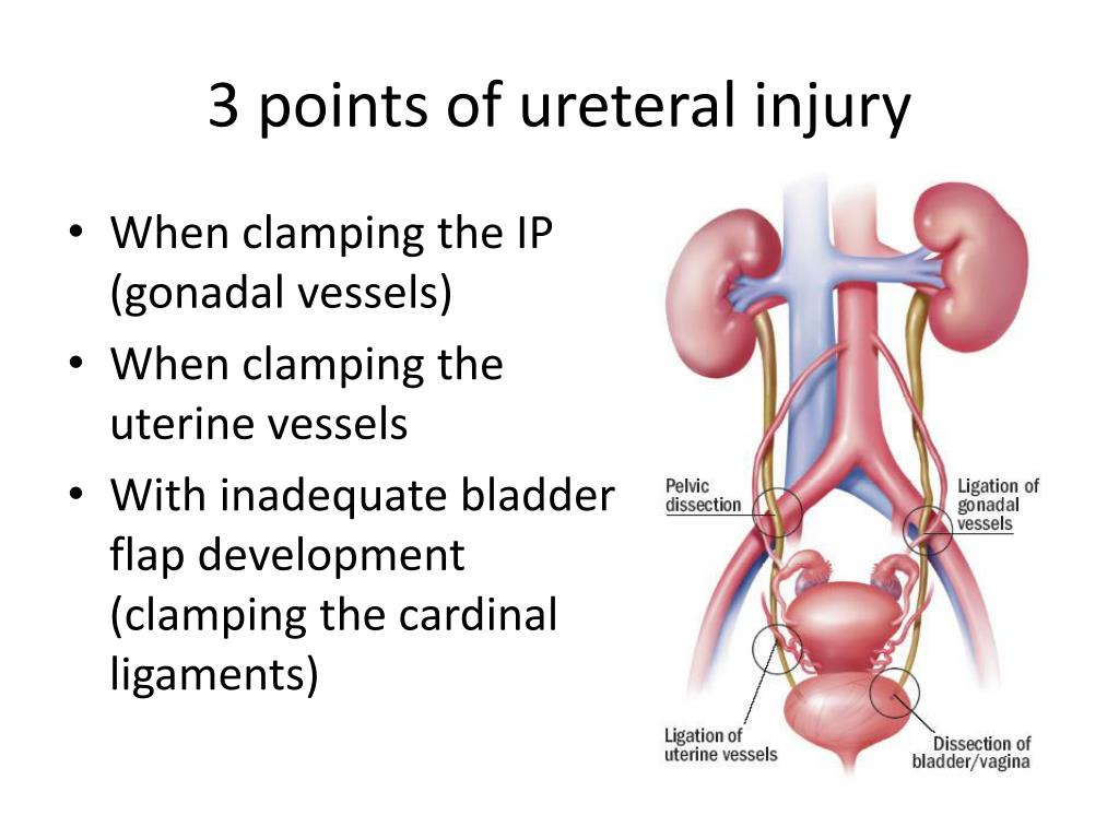
PPT Pelvic Surgical Anatomy PowerPoint Presentation, free download

Female Pelvic Anatomy Ureter ANATOMY STRUCTURE
In General The Ureter Is Seen Crossing The External Iliac Vessels From Lateral To.
It Begins At The Neck Of The Bladder, Traverses The Pelvic And.
Then, At The Level Of The Ischial Spine, The Uterine Artery Crosses The Ureter.
In The Pelvis, They Receive Additional Branches From The Internal.
Related Post: